MIT engineers have invented a revolutionary MRI-based method for spotting bioluminescence in the brain. This enables detailed imaging of deep brain structures and improves our understanding of brain functions and gene expression.
MIT has developed a new MRI technique that can show bioluminescence deep in the brain. This provides fresh insights into how brain cells develop and communicate with each other.
Researchers often use glowing proteins to track tumor growth or measure changes in gene expression as cells differentiate.
Although this method works well for cells and some body tissues, it has been hard to use it to image deep brain structures due to light scattering too much before it can be detected.
MIT engineers have devised a new way to detect bioluminescence in the brain by modifying the brain's blood vessels to react to light. This reaction can be observed using MRI, allowing researchers to identify the source of light.
Alan Jasanoff, an MIT professor, explains that it's challenging to use optical tools in deep tissue, and the study aimed to find a way to image bioluminescent molecules in deep tissue with high resolution.
The new technique could allow researchers to explore the brain's inner workings in more detail than before.
Jasanoff, an associate investigator at MIT’s McGovern Institute for Brain Research, is the senior author of the study published in Nature Biomedical Engineering on May 10. Former MIT postdocs Robert Ohlendorf and Nan Li are the lead authors of the paper.A new method for detecting bioluminescence in the brain uses MRI and could help researchers explore the brain's inner workings in more detail than before.
The technique, developed at MIT, allows researchers to explore the inner workings of the brain in more detail than previously possible by detecting bioluminescence using MRI.
Detecting Light
Bioluminescent proteins are found in many organisms, including jellyfish and fireflies. Scientists use these proteins to label specific proteins or cells, whose glow can be detected by a luminometer. One of the proteins often used for this purpose is luciferase, which comes in a variety of forms that glow in different colors.
Researchers at Jasanoff’s lab focused on finding a way to detect luciferase deep within the brain by transforming the blood vessels of the brain into light detectors. They accomplished this by engineering the blood vessels to respond to light by dilating, in line with how a popular form of MRI images changes in blood flow in the brain.
Alan Jasanoff thought they could use the ability of functional MRI and other imaging techniques to image blood vessels to also image light, by making the blood vessels sensitive to light.
To make the blood vessels react to light, the researchers modified them to produce a bacterial protein called photoactivated adenylate cyclase (bPAC). Beggiatoa When exposed to light, this protein creates a molecule called cAMP, which causes blood vessels to widen. This change in blood vessels can be detected by MRI because it affects the magnetic properties of oxygenated and deoxygenated hemoglobin.
BPAC is triggered by blue light and is good at detecting light close by. The researchers delivered the gene for bPAC to the smooth muscle cells in blood vessels using a viral vector. This made blood vessels throughout a large area of the brain sensitive to light.
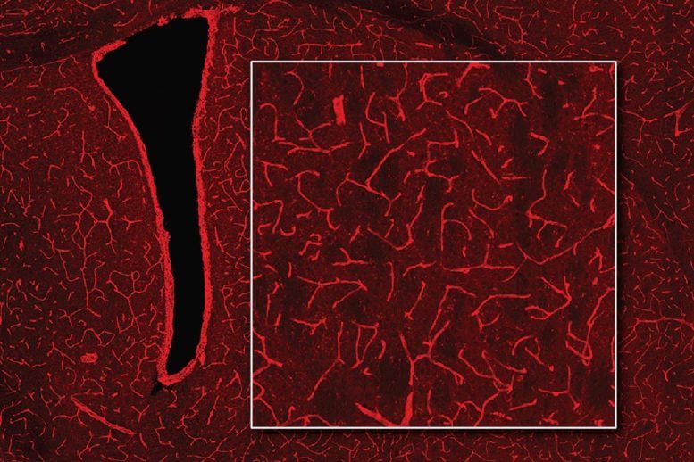
The brain has a dense network of blood vessels, so the researchers transformed the brain's blood vessel system into a three-dimensional camera.
Once the blood vessels became sensitive to light, the researchers implanted cells that were engineered to produce luciferase if a substance called CZT is present. When they imaged the brain with MRI, they were able to detect the dilated blood vessels by seeing the luciferase.
Monitoring Changes in the Brain
The researchers then checked if their method could detect light produced by the brain's own cells, engineered to produce luciferase. When the CZT substance was injected, MRI showed where light had been emitted.
The researchers call this method bioluminescence imaging using hemodynamics (BLUsH) and think it could be used in different ways to help scientists learn more about the brain.
One potential use is to track changes in gene expression by connecting the expression of luciferase to a specific gene. This could help observe how gene expression changes during embryonic development, cell differentiation, or when new memories form.
The researchers aim to explore these applications and adapt the technique for use in mice and other animals.
Reference: “Imaging bioluminescence by detecting localized haemodynamic contrast from photosensitized vasculature” by Robert Ohlendorf, Nan Li, Valerie Doan Phi Van, Miriam Schwalm, Yuting Ke, Miranda Dawson, Ying Jiang, Sayani Das, Brenna Stallings, Wen Ting Zheng and Alan Jasanoff, 10 May 2024, Nature Biomedical Engineering.
DOI: 10.1038/s41551-024-01210-w
The study was supported by the U.S. National Institutes of Health, the G. Harold and Leila Y. Mathers Foundation, Lore McGovern, Gardner Hendrie, Brendan Fikes, a grant from the German Research Foundation, a Marie Sklodowska-Curie Fellowship from the European Union, and a Y. Eva Tan Fellowship and a J. Douglas Tan Fellowship, both from the McGovern Institute for Brain Research.